Helaas is deze beschrijving nog niet in het Nederlands vertaald. U vindt hier dus een Engelse artikelbeschrijving.
Prepared Microscope Slides
Basic component of the program are the A, B, C and D series comprising of 175 microscope slides. The four series are arranged systematically and constructively compiled, so that each enlarges the subject line of the proceeding one. They contain slides of typical micro-organisms, of cell division and of embryonic developments as well as of tissues and organs of plants, animals and man. Each of the slides has been carefully selected on the basis of its instructional value. LIEDER prepared microscope slides are made in our laboratories under scientific control. They are the product of long experience in all spheres of preparation techniques. Microtome sections are cut by highly skilled staff, cutting technique and thickness of the sections are adjusted to the objects. Out of the large number of staining techniques we select those ensuring a clear and distinct differentiation of the important structures combined with best permanency of the staining. Generally, these are complicated multicolor stainings. LIEDER prepared microscope slides are delivered on best glasses with ground edges of the size 26 x 76 mm (1 x 3"). – Every prepared microscope slide is unique and individually crafted by our well-trained technicians under rigorous scientific control. We therefore wish to point out thatdelivered products may differ from the pictures in this catalog due to natural variation of the basic raw materials and applied preparation and staining methods.
The number of series in hand should correspond approximately to the number of microscopes to allow several students to examine the same prepared microscope slides at the same time. For this reason all slides out of the series can be ordered individually also. So, important microscope slides can be supplied for all students.
Parasites, pathogenic bacteria and insect pests for veterinary medicine, part IV
31 prepared microscope slides
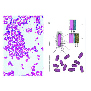
- Staphylococcus aureus, pus organism, smear from culture
- Hemolytic streptococci, blood poisoning, blood smear
- Bacillus anthracis, smear from culture
- Bacterium erysipelatos (Erysipelothrix rhusiopathiae), smear *
- Trichomonas sp., smear with trophozoites
- Plasmodium cathemerium, avian malaria, blood smear *
- Babesia canis, blood smear shows heavy infection
- Toxoplasma gondii, section of the brain showing cysts with parasites *
- Sarcocystis tenella in heart muscle, sec. showing Miescher's tubes
- Nosema apis, honey bee dysentery, sec. of diseased intestine
- Eimeria stiedae, causing coccidiosis in rabbit, section of liver shows schizogony and all developing stages
- Eimeria tenella, section of diseased chicken intestine *
- Dicrocoelium dendriticum (D. lanceolatum), sheep liver fluke, entire mount and stained for internal structures
- Fasciola hepatica, ova w.m.
- Taenia pisiformis, gravid proglottids w.m.
- Taenia pisiformis, ova from faeces w.m.
- Cysticercus bovis, bladderworm of Taenia saginata, sec. through beef muscle with parasites in situ
- Moniezia expansa, tapeworm of sheep, proglottids w.m.
- Dipylidium caninum, tapeworm of dogs and cats, immature proglottids w.m.
- Echinococcus granulosus, cyst wall and scolices t.s.
- Ascaris megalocephala, roundworm of horses, t.s. of adult female in region of sex organs
- Ascaris lumbricoides, ova in faeces w.m.
- Trichinella spiralis, section of infected muscle with encysted larvae
- Trichuris trichiura, whip worm, ova in faeces w.m.
- Onchocerca volvulus, sec. through host tissue with tumor containing larvae (filaria)
- Hirudo medicinalis, medicinal leech, t.s. through the body for demonstrating general structures of a leech
- Dermanyssus gallinae, chicken mite, w.m. *
- Varroa, parasitic mite of bees w.m.
- Ctenocephalus canis, male or female specimen w.m.
- Musca domestica, house fly, head and mouth parts with sucking tube, w.m.
- Culex pipiens, head and mouth parts of female w.m.
